Getting x-rays done at the dentist or orthodontist is a common part of many office visits. But did you know this technology wasn’t widely used for dental purposes until the 1950s?
Our team at Thacker Orthodontics wants to tell you how these x-rays work and, more importantly, answer the question, “Why do orthodontists use x-rays?” Let’s get started!
What Did We Do Before X-Rays?
Prior to wide access, traditional methods and clinical examinations were used. Orthodontists would still take photographs to document and track conditions during treatment, but obviously, that only relayed limited information. They also used something called panoramic radiographs, which were broader, less detailed images than modern x-rays.
Other methods included bite analysis and taking molds and impressions of the mouth, which could be used as a reference for the patient. They would also regularly take plaster models at different intervals to measure progress in the teeth and jaw.
In other words, it wasn’t quite as easy as it is now.
An Extensive Explanation
Dental x-rays are also known as radiographs. They are diagnostic tools that use electromagnetic radiation to capture pictures of teeth, bones, and all surrounding oral structures. Curious how they operate? Let’s break it down in detail:
- An X-ray machine generates controlled bursts of radiation. A dental assistant or radiologic tech will usually be operating this device.
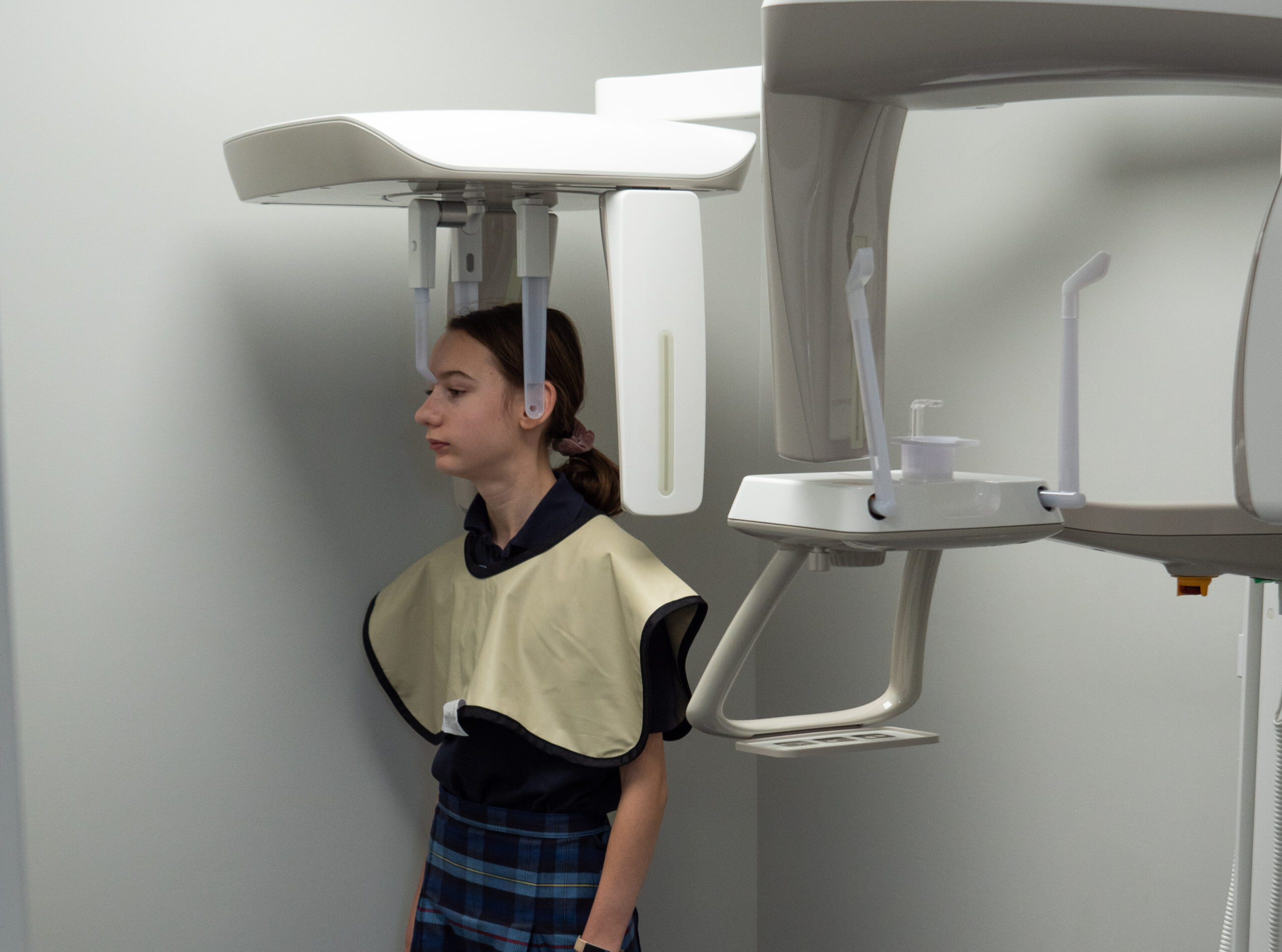
- You’ll be draped in something called a lead apron. It helps to block your vital organs from ionizing radiation. Usually, it will just cover your chest and torso.
- A digital sensor captures the images placed inside the patient’s mouth. It can be positioned in an area of interest, i.e., targeting a specific tooth. In the past, film was used for this purpose and would need to be developed in a lab.
- When the machine is activated, a focused beam of exposure is directed at the sensor. Teeth and bones absorb more x-rays due to their density, resulting in lighter areas on the x-ray image. Softer tissues like gums and cheeks allow more x-rays to pass through, creating darker areas in the image.
- The image appears on screen and is then examined by your doctor (more on that later!).
- Intraoral x-rays capture the inside of the mouth, helping make small areas visible.
- Extraoral x-rays capture the entire oral and facial structure, used for a broader view of the head and jaw.
Why Your Orthodontist Uses Them
Now that you know how it all goes down, we can talk about why and how Dr. B and Dr. MaryEvan utilize them!
- Assessment of Tooth and Jaw Position: The beauty of this technology is, first and foremost, that it provides us with images of the teeth, jawbone, and surrounding structures, which are not visible during a regular oral examination. These images aid us in assessing the position, alignment, and growth of teeth and jaws in patients of all ages.
- Early Diagnosis of Orthodontic Issues: We’re able to better track down orthodontic problems in their early stages—even before they become visibly apparent. This allows us to use interceptive treatment and potentially prevent issues from getting more severe.
- Identification of Impacted Teeth: When teeth are impacted, they haven’t erupted properly or are blocked from emerging. This information is vital for planning treatments, especially in cases where oral surgery is required. You might hear this phrase used when you’re getting your wisdom teeth removed (but hopefully not!).
- Evaluation of Dental Development: This technology helps us easily keep valuable records showing the development of teeth in children and adolescents. This comes in handy, particularly when observing how permanent teeth are coming in.
- Assessment of Bite Problems: X-rays can help orthodontists evaluate bite problems such as overbites, underbites, crossbites, and open bites (otherwise known as malocclusion). By visualizing the relationship between the upper and lower jaws and the alignment of teeth, we can better assess treatment options.
- Bone Health: Monitoring density and health of the jawbone is something you don’t always consider, but we do! When orthodontic treatment involves the repositioning of teeth, bones must be capable of supporting these changes.
- Treatment Verification: After the orthodontic treatment is complete, images can be taken to confirm that the teeth and jaws have been successfully aligned precisely according to the treatment plan.
Excellence Without Exceptions!
X-rays are just one of many tools we use as professionals to help us give you the best care possible. Thacker Orthodontics is committed to serving the communities of Cincinnati and Hillsboro with pride! Book your free consultation now—we can’t wait to meet you!